HISTOPATHOLOGY SERVICES
AAD Pathology is proud to have a dedicated in-house histopathology department, providing comprehensive diagnostic services for a wide range of biopsy submissions.
We are aware of our responsibilities to our partners and to human health, therefore we are continuously optimizing our processes and quality controls. Accuracy and precision are key for all our operational personnel and reflect the quality of our work.
In addition to reliability of our results, we consider discretion and loyalty as the most important principles in professional ethics of AAD Pathology. We act in an open and loyal manner towards our business partners and provide absolute confidentiality for our physicians and patients .
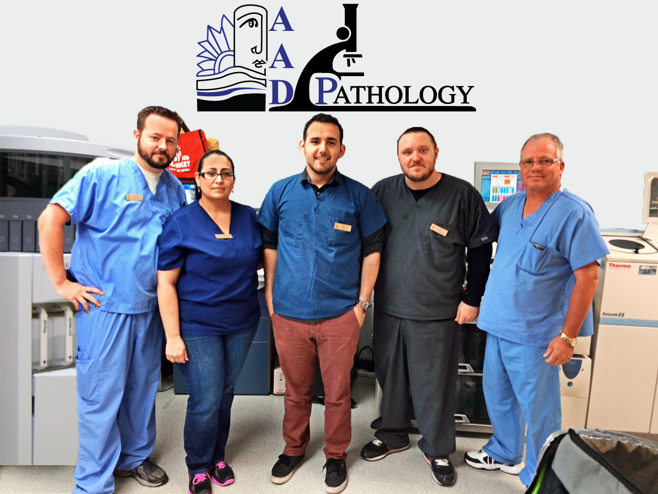
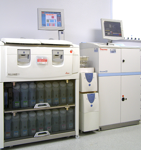
Our high throughput is owed to the years of experience we possess in the field of clinical histology. Our team is working in shifts. Qualified first-class equipment and optimized processes are the pillars of our success.
We take advantage of these privileges and continue to improve our professional service.
Lucy Soto
Pathology Laboratory Manager
Our Location
Located In Beautiful Tampa Bay Area
5210 Webb Road Tampa, FL 33615
Phone: (813) 882-9986 ext 1116 & 1137
Fax: (813) 885-5015
Email: info@AADPathology.com
Web: AADPathology.com
Slide Preparation

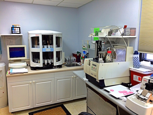
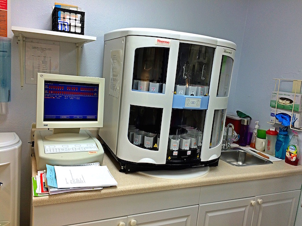
The specimens are accessioned by giving them a number that will identify each specimen for each patient and documented on a request form. Gross examination consists of describing the specimen and placing all or parts of it into a small plastic cassette which holds the tissue while it is being processed to a paraffin block. Initially, the cassettes are placed into a fixative.

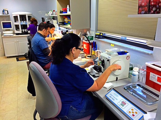
Once the tissue is paraffin infiltrated, it is embedded in a block of paraffin. The tissue sample is placed in a small mold that is then filled with melted paraffin. Once the paraffin has cooled and hardened, the mold is removed, exposing a small paraffin block containing the tissue sample.

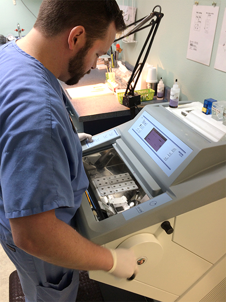
The paraffin block containing the tissue sample is sliced, or sectioned using a device known as a microtome. The microtome is equipped with a blade sharp enough to routinely slice the paraffin block into sections as thin as 2 micrometers. Once produced, the paraffin sections are floated on a water bath, from which they are mounted on glass slides.

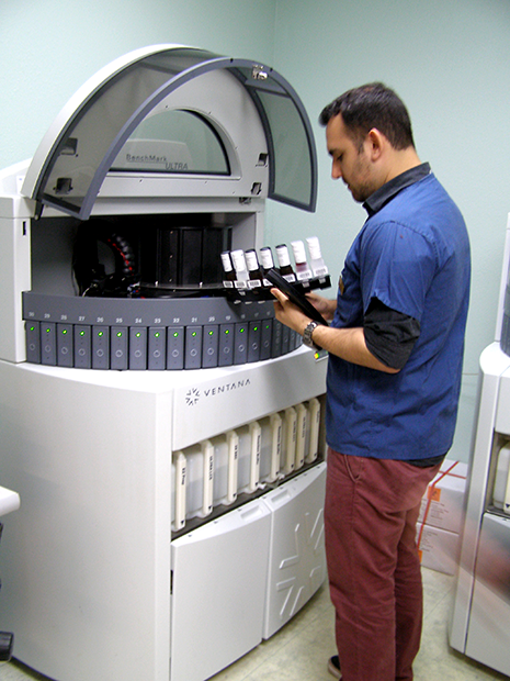
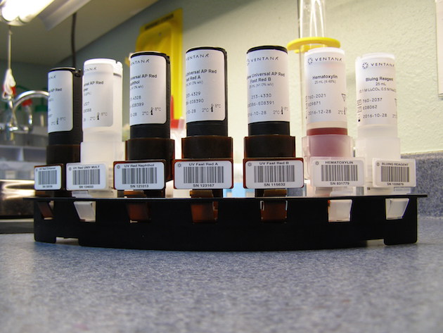
Once the tissue sections are mounted on glass slides, the sections must be stained to produce the contrast needed to visualize the tissue with optical microscopy. A classic staining protocol involves passing the slides through a series of baths containing a series of reagents that dissolve and remove the paraffin and properly stain the sections.

The resultant slides, together with the request forms are issued to the pathologist in the relevant subspecialty for rapid diagnosis. Digital pathology images are available on request.


