
Mohs Micrographic Surgery
Mohs surgery is a highly effective, specialized technique for removing skin cancers. The technique was developed in the 1930s by Dr. Frederick Mohs at the University of Wisconsin and is now practiced throughout the world. Mohs surgery differs from other forms of skin cancer treatment in that it allows for the immediate and complete microscopic examination of the removed cancer tissue so that all roots and extensions of the cancer can be eliminated. Mohs surgery has the highest reported cure rate of all treatments for skin cancer.
Treating all skin cancers with Mohs surgery is not necessary. Mohs surgery is reserved for skin cancers that grow back after previous treatment, cancers that are at high risk of recurring or for cancers that are located in cosmetic areas where preservation of the maximum amount of normal skin is important.
More than 3.5 million skin cancers in more than 2 million people are diagnosed annually. Our goal is to provide expert skin care with a gentle, personal touch. We are proud of what we do and of our focus on each patient.
Because this method requires doctors who have highly specialized training, dedicated personnel and specific surgical facilities, few medical centers are equipped to offer such treatment. In addition to the physicians, the team includes nurses who are experienced in caring for patients who have skin cancers at all stages of their diagnosis and treatment. Another important member of the team is a technician who quickly and skillfully prepares the tissue for microscopic examination. Finally, a patient care co-ordinator rounds out the team and is best qualified to handle your questions regarding insurance forms or other financial matters.
What kind of skin cancers can Mohs micrographic surgery be used for?
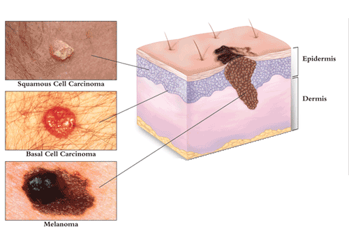
The most common types of skin cancer to be treated by Mohs micrographic surgery are Basal Cell Carcinoma (BCC) and Squamous Cell Carcinoma (SCC). Both types enlarge from the point where they first occur and usually do not spread (metastasize) to distant parts of the body. If not completely removed, both will invade and destroy structures in their paths. Compared to other forms of cancer, these types of skin cancer can be recognized in their early stages and are therefore easily cured.
How does skin cancer grow?
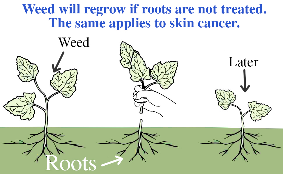
It is important to understand how skin cancers grow, as this in turn will help appreciate why Mohs micrographic surgery is so useful. Skin cancers begin in the uppermost layer of the skin (the epidermis) and grow to the sides on the surface of the skin and downward, below the skin surface, with root-like extensions. This is comparable to the roots of a weed. Unfortunately, root-like extensions of skin cancer cannot be seen with the naked eye. Therefore, what is apparent to the naked eye on the surface of the skin may actually be only the visible ‘tip of the iceberg’. If the roots are not treated, then the skin cancer will likely recur. Mohs micrographic surgery helps ensure these roots are cleared away completely as removed tissue is examined under the microscope and the tumor is mapped so that guessing the extent of the tumor is eliminated. All other methods of skin cancer treatments require best guessing how wide and deep to treat.
How is Mohs micrographic surgery performed?
Mohs micrographic surgery is performed as a day case procedure, usually under local anesthetic. This method involves the steps below:
The Procedure
The Mohs surgical process involves a repeated series of surgical excisions followed by microscopic examination of the tissue to assess if any tumor cells remain. Some tumors that appear small on clinical exam may have extensive invasion underneath normal appearing skin, resulting in a larger surgical defect than would be expected. It is therefore impossible to predict a final size until all surgery is complete. As Mohs surgery is used to treat complex skin cancers, approximately half of all treated tumors require 2 or more stages for complete excision.
Our Mohs surgeons at Academic Alliance in Dermatology remove the cancer layer by layer, examining each one microscopically until the margin around the cancer is free of cancer cells. This is a technically precise process in which the physician serves both as the surgeon and pathologist.
Anesthesia
The tumor site is locally infused with anesthesia to completely numb the tissue. General anesthesia is not required for Mohs micrographic surgery.
Removal of visible tumor
Once the area has been completely numbed, the tumor is gently scraped with a curette, a semi-sharp, scoop-shaped instrument. This helps define the clinical margin between tumor cells and healthy tissue. The first thin, “layer” of tissue is then surgically removed by the Mohs surgeon. An electric needle may be used to stop the bleeding.
Mapping and Tissue Processing
Once a “layer” of tissue has been removed, the patient returns to the waiting room. The tissue is processed for approximately one hour. This process begins with a “map” or drawing of the tissue and its orientation. The tissue is color-coded in 4 different colors to denote top from bottom and left from right. These colors are documented on the map to serve as a guide to the precise location of any cancerous cells, if present.
The tissue is frozen and cut into thin pieces 1/10 the thickness of a piece of paper.
The thin pieces of tissue are placed on a slide and stained so that cancer cells are visible under microscope.
Examination
Our Mohs surgeon examines the slides under a microscope.
The entire undersurface and skin edges of the removed tissue are evaluated microscopically by our Mohs surgeon to determine if the cancer has been completely removed. If cancer remains, the Mohs surgeon marks on the map where the tumor is still present and another layer of tissue is removed and examined.
Second Stage
If any skin cancer remains, more tissue is removed using the map as a guide. Due to the “mapping” technique, the surgeon is able to remove only the cancer and preserve healthy tissue keeping the wound as small as possible. The tissue is processed and examined under the microscope. This process is repeated until all cancer cells are removed.
Reconstruction
At Academic Alliance in Dermatology our Mohs surgeons are experts in the reconstruction of skin defects. Reconstruction is individualized to preserve normal function and maximize aesthetic outcome. The best method of repairing the wound following surgery is determined only after the cancer is completely removed, as the final defect cannot be predicted prior to surgery. Stitches may be used to close the wound side-to-side, or a skin graft or a flap may be designed. Sometimes, a wound may be allowed to heal naturally.
More Questions:
How long does the procedure take?
Most cases can be completed in less than four stages, requiring less than four hours. However, no one can predict how extensive a cancer will be because the size of a skin cancer’s roots can’t be estimated in advance. Therefore, we ask that you reserve the entire day for surgery in case additional surgical sessions are required.
Will the procedure leave a scar?
Yes, any form of surgery leaves a scar. However, the Mohs micrographic surgical procedure will leave one of the smallest possible surgical scars.
What happens after the Mohs surgery is completed?
After the cancer is removed, our Mohs surgeons will discuss options with you. These options may include allowing the wound to heal naturally, without additional surgery (often produces the best cometic results), or having the wound repaired by one of the physicians.
Will I have pain, bruising or swelling after surgery?
Most patients do not complain of significant pain. If there is discomfort, Tylenol is usually all that is necessary for relief.
However, a stronger pain medication will be prescribed if needed. You may have some bruising and swelling around the wound, especially if surgery is being performed close to the eyes.
Will my insurance cover the cost of Mohs?
Most insurance polices cover the cost of Mohs surgery and the surgical reconstruction of the wound. The staff at Academic Alliance in Dermatology will confirm that your insurance company will pay for your procedure prior to your surgery.
How do I prepare for the surgery?
Please get a good nights rest and eat normally the day of the surgery. If you are taking prescription medications, continue to take them unless otherwise directed. You may want to bring a book/magazine with you to occupy your time while waiting for your slides to be processed and examined.