
DEPARTMENT OF DERMATOPATHOLOGY
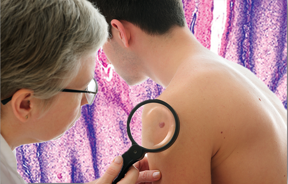 Our dermatopathology laboratory is known as a center of excellence for dermatological specimens. It is critical to the Academic Alliance in Dermatology team to interpret and deliver accurate, timely results for the best possible patient care. With diagnostic services covering a broad spectrum of dermatology disorders, we share in our client’s focus on high quality patient service.
State-Of-The-Art
What We Offer
Biopsies sent to our lab are processed in our state-of-the-art laboratory, overseen by our quality assurance program. For those cases that require more sophisticated tests, we can offer a comprehensive variety of immunostains, dual immunostains, special stains and direct immunofluorescence. All cases are interpreted by our board certified dermatopathologists, Dr. Iriana Belongie, MD, FAAD and Dr. Charles Knapp, MD, FAAD.
EMR (Electronic Medical Records)
At our facility we are in a unique position as all relevant medical data and clinical pictures are available to our dermatopathologists through our electronic medical records. This enables clinical pathological correlation which enhances the quality of our care. Our rich and broad patient population exposes us to patients of all ages and many different ethnicities. This makes the practice of dermatopathology both unique and exciting.
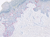
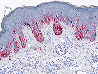
Inflammatory and other skin diseases may require special staining to detect certain cell types or infectious agents such as fungi and mycobacterium. The majority of skin biopsies and excisions are successfully analyzed using routine histopathology (H&E). Special stains and ancillary testing are ordered only when necessary or when requested by the clinician.
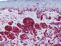
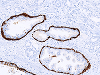
A full range of antibodies against tissue and cellular biomarkers are available for evaluation of neoplasms including epithelial tumors, melanocytic lesions, lymphoproliferative malignancies and poorly differentiated spindle cell tumors.
- Pemphigus and Pemphigoid Groups
- Dermatitis Herpetiformis
- Lupus Erythematosus & other Collagen Vascular Diseases
- Vasculitis versus Urticaria
- Porphyria Cutanea Tarda
- Pigmented lesions (melanoma)
- Metastatic neoplasms
- Soft tissue neoplasms
- Vulvar biopsies
- Primary cutaneous neoplasms (basal cell, squamous cell carcinomas)
- Adnexal neoplasms
- Inflammatory skin diseases (including blistering diseases, lupus erythematosus, vasculitis)
- Nail disorders
- Alopecia
Mohs Micrographic Surgery
Advantages of Mohs Micrographic Surgery
- Mohs surgery has the highest cure rate for skin cancer.
- It is especially suited for poorly defined tumors, recurrent tumors and tumors known to behave in an aggressive manner.
- Mohs surgery provides the surgeon and patient with confidence that the tumor is completely removed prior to closure of the wound.
- With Mohs surgery, less adjacent normal skin is sacrificed resulting in smaller wounds and therefore smaller scars.
Special Histology Test Menu
- Routine Biopsies
- Histochemical and Immunohistochemical Staining
- Direct Immunofluorescence Testing
- Mohs Micrographic Surgery